Encouraging patients to participate in their dental care is not easy, so I like to educate them with technologies that offer a visual representation. For example, when patients can see dental caries on their teeth, they understand why I recommend certain restorative procedures. DEXIS CariVu™ caries detection device offers a no-radiation, highly accurate, visual option for caries detection and patient education.
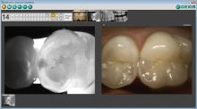
CariVu gives an impressive visual image of caries similar to an X-ray. The enamel appears transparent, and caries shows up as dark areas that are easy to see and understand. CariVu, with its broad use for occlusal, interproximal, and recurrent carious lesions and cracks, offers 99% accuracy. We use it on any patient not yet due for radiographs or who is reluctant to allow radiographs. In some cases, after we use CariVu, the patients are more willing to allow radiographs to further determine if treatment is necessary.
During capture, DEXIS CariVu images are automatically compared with X-rays and photos.
When patients arrive in the hygiene room, we discuss normal office protocols, like frequency of radiographs. The hygienist will capture radiographs and may capture CariVu images. Even with radiographs, we use CariVu to double-check caries that may not yet be visible on the X-ray. If a patient asks about the images, the hygienist educates the patient on proper dental health and care because CariVu images can be a good illustration of good and not-so-good hygiene.
“Our digital radiography, intraoral camera, and CariVu create a whole package that makes a huge difference for our patients’ comprehensive dental care.”
As part of our office technology, monitors are positioned above the patient’s head, and we can show CariVu images and explain our findings. It is easy to see the difference between the perfect tooth and a tooth with a carious lesion.
As a prosthodontist, implants and crown preps are a growing part of my practice, so it is very important to discover if caries exists between teeth. If I see a suspicious area, I snap an image with CariVu to document it and monitor changes over time. As a result, we only do restorative procedures when they are needed — when the caries indicate treatment— and not before, and patients appreciate that.
I know that CariVu is making a positive impression when patients return and ask if we can use “the light device” again. They like to see whether they can tell if a carious lesion has progressed or not—the image is that definable. Our digital radiography, intraoral camera, and CariVu create a whole package that makes a huge difference for our patients’ comprehensive dental care.
This article was originally published in Sidekick Magazine.

About the Author: Dr. Rich Stuart received his undergraduate degree from Ball State University and his dental degree and post-graduate training in prosthodontics from Indiana University School of Dentistry. He is a proponent of organized dentistry and a Fellow of the International Team of Implantologists. Dr. Stuart is the team dentist for the NFL’s Indianapolis Colts and the Indianapolis Ice hockey players, and he maintains a private practice in the city.