Cone Beam Technology Offers Enhanced Diagnostic Capabilities and Improved Treatment Planning With Excellent Imaging Quality.
About 3 years ago, I invested in a 2D pan machine, and we used it routinely. However, my decision to start placing implants was the tipping point for upgrading to cone beam 3D imaging. Although it’s not a requirement for placing implants, a 3D image is such a valuable piece of information, and I thought it worth the investment to get a unit here in the office so I could thoroughly plan out the entire implant procedure and minimize or eliminate any surprises.
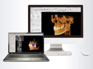
Invivo 5.4 and Invivo 5 for Mac are powerful CBCT 3D imaging applications specialized for implants, orthodontics, oral surgery, and restorative dentistry. Both offer fast, quality rendering and visualization on cone beam 3D scans and tools for accurate treatment plans.
After considering my options, I realized that KaVo was the best choice based on a number of factors. It seemed to have the best integration with DEXIS software, the price was in the range I was looking for, along with the features I was looking for, and the size of the unit was right based on the space I have.
While the driving force for clinical use is implants, we use 3D imaging quite a bit as a diagnostic tool when trying to determine what’s really going on with the tooth and to help guide a proper course of action. For instance, we use it whenever a patient complains about a toothache or exhibits other signs or symptoms, and we can’t definitively discern what the issue is with standard 2D x-rays. 3D imaging enables us to pick up small fractures or detect a root fracture or a missed canal on a root canal.
Getting Acquainted
In the beginning, the actual cone beam scan took longer. As you do additional scans, though, you get a better feel for adjusting the 3D unit’s settings, so it really isn’t that much longer than taking a 2D pan. There’s a little bit of a learning curve to viewing a 3D image, in the same way a 2D x-ray can be a little confusing. It takes some time to get the orientation, but after you look at several scans, especially with some professional guidance, you become an expert, and at a certain point, you can look at them without thinking twice. It’s similar with 3D because when you first look at a scan on the computer screen, it can seem a bit daunting.
I took a CE course at the University of Minnesota that walked me through the software and different settings and viewing angles, and that was very helpful. I certainly encourage dentists to take a course on how to manipulate the images and know what you’re looking at.
In addition, we had a full day of training with KaVo to get up and running with the software. There are also many online tutorial videos, and you can probably learn everything you need to know just by reviewing those. The trainer has been available, too, if I have any specific questions.
GOING GREEN WITH OP 3D
The OP 3D’s versatile panoramic and 3D programs offer imaging excellence for a variety of uses, including implantology, endodontics, TMJ, airway, trauma, and periodontics. It’s also a sustainable green solution because it replaces lead typically used for tubehead radiation shielding designs with a more ecological and environmentally friendly alternative.
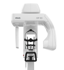
Special features include:
• ORTHOselect – Optimizes workflow by enabling the user to make selections of individual teeth, an entire upper or lower jaw, or TMJ.
• Customized FOVs with SMARTVIEW 2.0 – Enables the user to choose the most optimum field-of-view size based on the clinical need.
• QUICKcompose – Offers a quick preview of the captured image allowing for timely evaluation.
The OP 3D offers 4 versatile FOVs:
• 5 cm x 5 cm • 6 cm x 9 cm • 9 cm x 11 cm • 9 cm x 14 cm
The Clinical Difference
Aside from placing implants, I think the biggest benefit of using 3D is that it allows us to arrive at a diagnosis much more quickly in situations where the standard visual exam and 2D x-ray exam are inconclusive. We can get the patient proper treatment more quickly.
In a recent case, we noticed on a regular 2D pan that there were some unusual objects in the patient’s left TMJ. So we took a 3D scan of both the left and the right TMJ. I looked at the results myself and also sent the scans to the University of Minnesota Oral Radiology Department so they could do an evaluation. With this confirmation, I was able to properly diagnose the patient and plan a course of treatment.
In a world where 3D technology is impacting the games our children play and the movies we watch, it’s important for dentists to harness the benefits of 3D imaging. It allows us to visualize our patients’ teeth and jaws with better clarity than ever before, and as a result, we can diagnose better and provide treatment plans that are more accurate, timely, and cost-effective.
CLOSE-UP ON OP 3D PRO
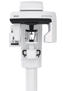 Available in four upgradable models and offering traditional panoramic and optional cephalometric imaging, the OP 3D Pro is KaVo’s most comprehensive 3-in- 1 unit for the entire maxillofacial region. The OP 3D Pro’s easy-to-use, 10-inch user interface enables intuitive usage and setting of imaging parameters from the very beginning. It can be used for all clinical needs, including general practitioners, endodontics, implantology, orthodontics, oral and maxillofacial surgery, periodontics, prosthodontics, and airway.
Special features include:
• SMARTVIEW functionality – Allows FOV positioning accuracy to be verified or adjusted if needed before CBCT examination.
• Automatic Dose Control for 3D – Obtains patient-specific exposure settings to provide premium-quality images at optimal dose for the patient.
• Multilayer pan – Provides 5 panoramic images with only one scan to compensate for incorrect patient positioning and difficult anatomies, all achieved in the same scanning time and dose as the traditional panoramic scan.
This article originally published in Dental Product Shopper – January 2018, and later published in Sidekick Magazine.
 About the Author: Dr. McGann graduated from the University of Minnesota School of Dentistry in 2002 and completed a residency at the Minneapolis Veterans Administration Medical Center with a focus in oral surgery. He is an alumni member of the Dawson Academy, which provides instruction on complex restorative and esthetic cases. Dr. McGann is an adjunct professor at his alma mater and enjoys working with students at outreach clinics. He volunteers at the Union Gospel Mission, St. Paul Dental Connect, Minnesota Mission of Mercy, Twins and Vikings Team Smile, and Give Kids a Smile, and has traveled to Uganda on 3 occasions to treat children who would otherwise receive no dental care.