Many endodontists are curious about cone beam computed tomography (CBCT) and what it would be like to use this advanced imaging technology in their practices. Often, their questions are practical ones: What is the importance of CBCT? When should it be used: preoperatively, intraoperatively and postoperatively? Are all CBCT units the same? How do you read a CBCT scan? How do you interpret the results?
To shed some light on what it’s like to use CBCT in daily practice, Dr. Stephanie Tran, an endodontist with a private practice in New York City and Long Island, New York, offered insights based on her own use of CBCT. Dr. Tran finished her undergraduate and doctoral studies at the University of the Pacific in San Francisco, California, and served as chief resident at the University of Tennessee Health Science Center, where she completed her post-graduate endodontics specialty residency.
Why 3D imaging can be helpful, in conjunction with 2D images, and when it is used
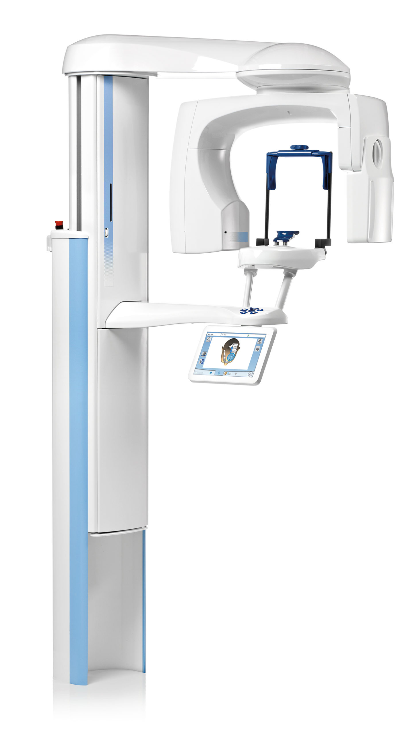
The 3D images dentists can generate using CBCT provide a more comprehensive view of the mouth than 2D versions. Understanding how to take high-quality 3D images is essential to getting the most benefit from this technology. Since each type of CBCT machine has its own features and functionality, it is important to schedule a demonstration and training session with the manufacturer of any machine you are considering before you purchase it, to make sure you understand what it can do and that you are comfortable using it. Machines vary considerably when it comes to capabilities, such as resolution and field of view.
During the consideration process, many dentists often wonder how much radiation exposure patients will undergo when using CBCT. Fortunately, CBCT units with a local, limited field of view have very minimal radiation. This is an attractive feature, particularly for endodontists, given that it is very important to take 3D images for diagnostic and treatment planning purposes.
That said, a CBCT unit should not be used for routine screenings. Prior to doing a CBCT scan, an endodontist will generally want to use simpler diagnostic methods, such as a 2D radiograph. This will allow a perspective on what kind of bone loss there is, vertical as well as horizontal; where the bone loss is located, such as apically, mid-root or in the alveolar ridge; any furcation involvement, furcation bone loss, existing caries and restorations; and the shape and presence of any lesions, radiopacities (the ability of materials to block the passage of X-rays) and radiolucencies (radiographic changes indicating abnormalities). There can be false positives with periapical radiolucencies when using CBCT, so a 2D radiograph is better to use at this stage.
Armed with this information, the endodontist may decide a more detailed view of the mouth is necessary and opt for a CBCT scan. The endodontist will then make a restorative diagnosis, periodontal disease diagnosis and endodontic diagnosis. “CBCT is just one piece of the puzzle,” says Dr. Tran. “We still want to understand the patient’s symptoms and what we find clinically — as well as the 2D radiographs — to help us make that diagnosis.”
As Dr. Tran points out, there is an art and science to treating patients. “Endodontics involves the science of understanding the inside of the tooth, of understanding microbiology and of figuring out the mystery of the diagnosis,” she explains. “There’s also an art to it — the artistry of delicately treating the inside, while being minimally invasive. The end result of putting the right equipment to work for a practice is a patient experience that is ‘comfortable, relaxing and even fun.’”
Methods for obtaining CBCT images
Getting training from the manufacturer on how to use the right magnification is essential for getting the most out of CBCT scans, facilitating better diagnostics and patient education. Here are some of the key parts of the process that are important to master:
Field of view. Just as in 2D radiographs, images with the highest resolution are the clearest and easiest to read accurately. The same goes for 3D imaging, where the resolution must be extremely high to allow for accurate reading. The field of view size and resolution are inversely related. The larger the field of view and the more structures that are included, the poorer the resolution is. The smallest fields of view are going to give the highest resolutions, as well as the most information about a very, very small area. Keep in mind that the larger the field of view, the larger the quantity of information you must read and interpret. And the smaller the field of view, the higher the quality of information — it will be far more detailed and informative, with less material that requires a dentist’s interpretation.
Getting training from the manufacturer on how to use the right magnification is essential for getting the most out of CBCT scans.
Location. Location influences the field of view. Focusing the center of the CBCT image and taking the scan specifically on the tooth or couple of teeth in question is important. Make sure to include vital structures in the image, so you can see where they are in relation to the tooth.
“One important tip is to set the CBCT sometimes to be centered away from the restoration itself,” says Dr. Tran. “So, for example, crowns and any kind of restorations will cause artifacts in the image. Sometimes you can actually choose to center it more apically, or more mid-root, and actually cut out most of the restoration from the image.”
Avoiding restorations in the image allows you to avoid beam-hardening artifacts, which can sometimes make it easier to read the CBCT scan.
Voxel size minimums for the machine. If the machine can use very, very small voxels (a unit of graphical information), then the smaller they are, the clearer the image is going to be. The smaller field of view will be more focused. Different machines have different capacities when it comes to slicing and voxel sizes.
Kilovoltage peak (kVp) and milliampere-seconds (mAs). The clarity of your image will be determined by what kind of machine you have, what kind of field of views it can take and what kind of adjustments you can make for the kVp or mAs. Some machines will let you change or control all the kVp, mAs and radiation levels. So, for example, some machines will take an initial pan for the patient. The dentist clicks on the tooth, and the machine will automatically center itself on that tooth. Some machines have small laser guides that can be focused on the tooth itself. With regard to the very small field of view that’s required in endodontics, these guides are the equivalent of a sextant.
External factors. The computer screen itself and the lighting in the room can affect the quality of a CBCT image. While a dark room is ideal, says Dr. Tran, “just turning off the room lights helps a lot.” Beyond this, your software itself will definitely affect the clarity of the images. “Software that is specifically meant for small field of view or high-resolution images is going to be very important,” says Dr. Tran. Manipulating the image once you’re in the viewing software can also make it clearer. Some programs, and even the CBCT machines themselves, come with filters that can affect factors like what whites and darks look like. You can also set how many slices you want to take, where the slices are taken and the thickness of the slices after you’ve taken the scan.
What to look for when you review a CBCT scan
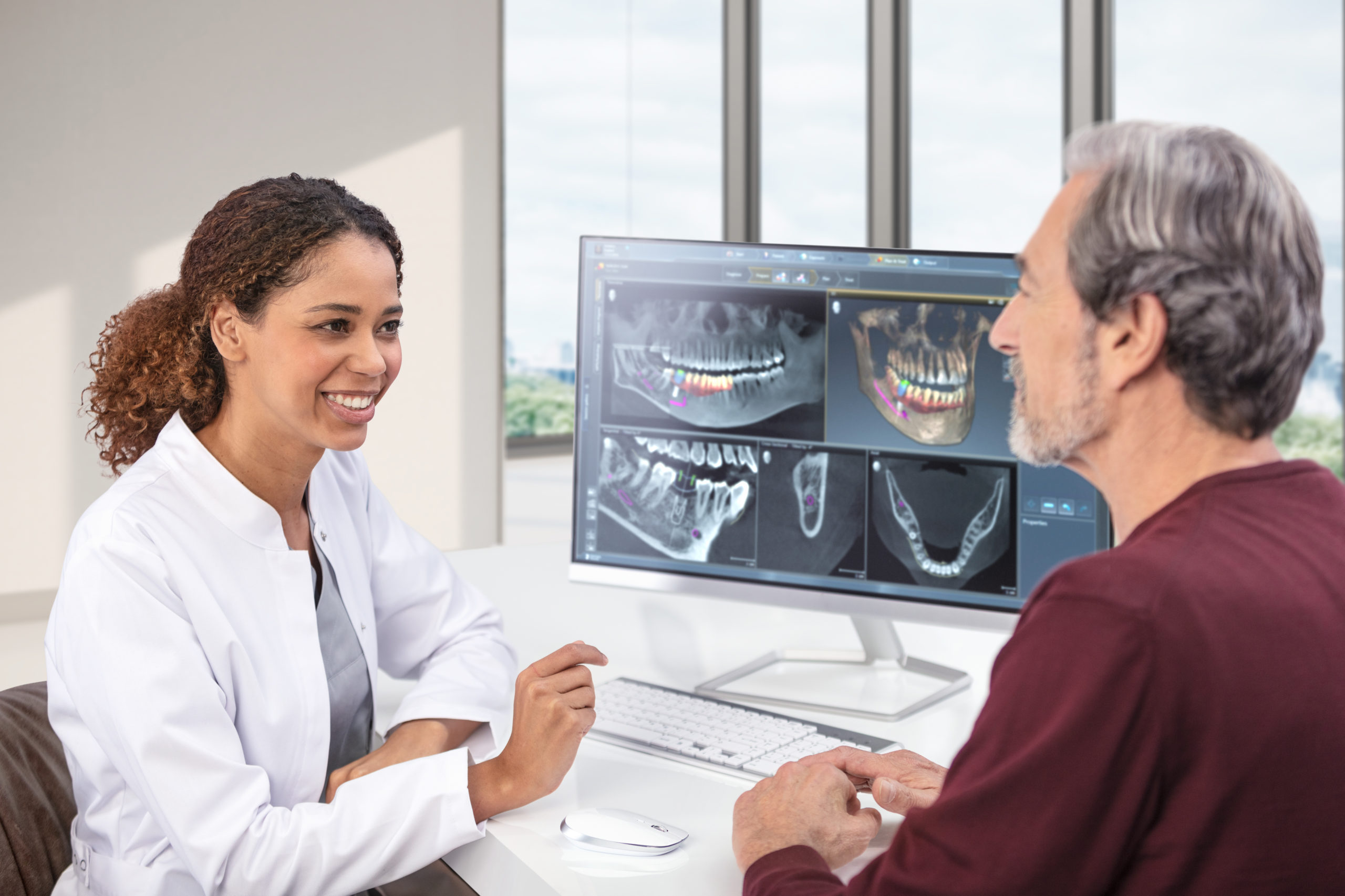
When reviewing a CBCT scan, there is a great deal of information to look at. “You’re looking at all structures around the tooth, such as the bone, the periodontal ligament and the lamina dura. You’re also looking at what has thickening or widening, what has bone loss, where the bone loss is and the extent of it. For example, is the bone loss apical, mid-root or at the ridge?” says Dr. Tran. “And then, are there radiolucencies or radiopacities? If there is an existing root filling, does it extend all the way down the canal? Are there any missed canals? How close is that root filling to the rest of the root structure? Is it too close to a wall? What kind of vital structures are nearby? And how close are they? What does the anatomy look like of both the tooth, the root and the bone, as well as the possibility of any root fractures?”
All of this can sound overwhelming at first, but, says Dr. Tran, “It’s just like when you’re learning how to read a 2D image. It’s about getting used to reading a 3D image, and understanding what to look for and what to evaluate.”
Interpreting the results of a CBCT scan
Once you know what you’re looking for, it’s time to evaluate what you’re seeing. Here are some key factors to keep in mind:
- What vital anatomical structures are near the tooth, and which are in the distance? An endodontist will want to know how the anatomical structures are behaving in relation to the tooth. For instance, you’ll want to look for mucosal thickening in the lining of the sinus, a sign that inflammation is about to occur. Tooth inflammation can sometimes manifest as a sinus reaction. Similarly, sinus issues can sometimes show up as odontogenic issues, such as jaw pain.
- The anatomy of the tooth itself. 2D images don’t always show the apex of a curvature of a tooth. This is because of the overlap of the different structures.
- The completeness of a root canal treatment. A 3D image can show if a root canal treatment is centered and if there are any missed canals. The canal should be centered within the root.
- The severity of non-odontogenic lesions. Clinical findings, 2D images and the results of tests such as the sensitivity of teeth to cold must also be considered to make a correct diagnosis.
A CBCT scan can also be used for surgical treatment planning. When considering an apical or external resorption defect treatment, one of the most important aspects of a CBCT scan is the measurements. Measurements taken on a CBCT scan are actually extremely accurate. “You don’t have that foreshortening issue that you have with a 2D radiograph,” says Dr. Tran.
Conclusion
Endodontic cases can be extremely complex and require dentists to use all of the tools at their disposal — from their clinical knowledge and expertise to the right imaging technology — to make a correct diagnosis. A CBCT machine, with its advanced 3D imaging capabilities, can be an important asset in any practice’s toolkit.
To explore other topics related to dentistry, dental equipment and dental practice solutions, visit https://henryscheinequipmentcatalog.com/content-library.