Digital imaging is part of efficient communication, and it adds to the “wow factor.” Patients may see their teeth in the mirror every day, but we must supply images that will let them learn about improving their dentition. For this, digital radiography has become the standard of care in diagnostics. This technology provides superior-quality images in a couple of seconds. It is an excellent diagnostic aid with which to compose a treatment plan. On the screen, we can enhance or zoom in, invert or emboss an image to communicate the possibilities in an unobtrusive way.
Sometimes a patient may have a problem such as a fractured restoration that cannot be seen or felt. In this situation, we call upon our Gendex digital technology collection: Digital intraoral and panoramic imaging, high resolution intraoral cameras, as well as my integrated patient education systems. All of these tools help the patient see situations that may not be clinically evident but certainly are present.
Among my other patient comforts are my digital sensors that capture intraoral digital X-rays; they have rounded corners that are more comfortable than traditional film. Digital sensors as a whole are excellent, but I made my selection based on comfort, portability, and its USB connectivity. It’s a plug and play device that travels all over my office, everyday.
In my practice, we utilize the panoramic as a screening tool. Our standard radiographic series consists of four bitewings and a panoramic X-ray. The resolution on digital panoramic radiographs are far superior to any image I have seen in my career, including film based images. Adding to the patient wow factor is the speed of acquisition. The 12 seconds it takes to capture the image makes digital panoramic imaging the most cost effective diagnostic revenue generator.
Instant, high-resolution digital X-rays–a practice essential
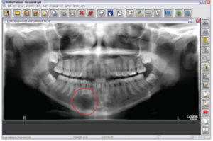
Besides aesthetics, the pan is also my first and foremost tool for general pathology. From the pan, I find lesions, supernumeraries, and situations that greatly decrease the morbidity and mortality of my patients. For example, a routine pan on a 17-year-old showed a 3-centimeter traumatic bone cyst that was not diagnosed from a set of intraoral X-rays. As a result, the patient had surgery, and was able to keep all of her teeth. If the cyst was not found and treated at that time, she could have experienced major loss of her mandible as well as teeth. Digital imaging technology has turned out to be life changing for me and for my patients.
Dr. William J. Bennett, III, maintains a private practice in Orland Park, IL, where he dedicates his career to providing his patients with healthy as well as beautiful smiles. Dr. Bennett graduated from University of Illinois at Chicago School of Dentistry in 1990, and since 1992, he has been on the Manor Health Care facility healthcare team in Palos Heights, caring for residents' dental geriatric needs. Dr. Bennett lectures nationally on technology in dentistry, as well as other topics. He is found on the Web at www.smilesbybennett.com.