- Home
- Digital Imaging
- Extraoral Imaging
- Planmeca ProMax® 3D Plus CBCT Unit
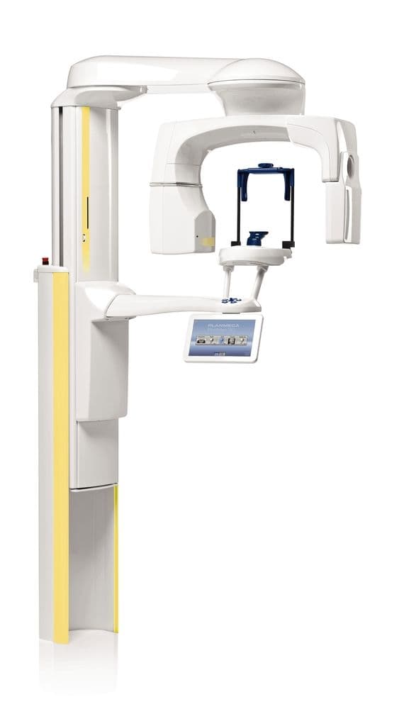
Planmeca ProMax® 3D Plus CBCT Unit
The Planmeca ProMax 3D Plus CBCT unit combines advanced 3D imaging capabilities with innovative features like movement correction and low-dose protocols, making it a trusted solution for general dentistry, endodontics and beyond.
Description
The Planmeca ProMax 3D Plus CBCT unit is a versatile and powerful 3D imaging tool that extends beyond standard dental applications, offering enhanced diagnostic capabilities for the entire maxillofacial region. Whether visualizing intricate sinus structures or capturing the smallest bone details in the ear, this CBCT unit provides exceptional image clarity and precision. Supporting panoramic, cephalometric and 3D imaging, the Planmeca ProMax 3D Plus features a dedicated endodontic mode that delivers high-resolution images of small anatomical structures, ensuring diagnostic accuracy in even the most detailed cases. Its Planmeca CALM® technology reduces motion artifacts, while Ultra Low Dose™ technology ensures minimal radiation exposure without compromising on image quality, offering a safer, more comfortable patient experience.
- SmartPan™ technology. The Planmeca 3D Plus features a unique system that allows seamless 2D and 3D imaging using the same sensor, streamlining workflow and reducing patient positioning time
- Universal accessibility. Designed with face-to-face patient positioning, accommodating patients in standing, sitting or wheelchair-bound positions for maximum comfort and accessibility
- SCARA technology. The patented Selectively Compliant Articulated Robotic Arm (SCARA) offers unrestricted movement during rotational imaging, ensuring highly accurate maxillofacial scans
- Versatile field of view. The Planmeca ProMax 3D Plus is capable of capturing a wide field of view (up to Ø20x10 cm) in a single scan, providing comprehensive imaging for diagnostic needs across multiple specialties
- Integrated workflow. Compatible with both Mac OS and PC systems, this CBCT unit integrates seamlessly with Planmeca Romexis®, a robust software platform that supports the entire CAD/CAM workflow, enhancing productivity and efficiency
Specifications
Anode voltage 60–90 kV, 60–120 kV
Anode current 1–14 mA
Focal spot 0.5 mm, fixed anode
Image detector Flat panel
Image acquisition 200 / 360-degree rotation
Scan time 9–33 s
Typical reconstruction time 2–30 s
Maximum volume sizes
Maximum volume with a single scan
Ø20 x 10 cm
Dental programs
Volume size (child mode) [cm]
Tooth
Ø4 x 5 (Ø3.4 x 4.2)
Ø4 x8 (Ø3.4 x 6.8)
Teeth
Ø8 x 5 (Ø6.8 x 4.2)
Ø8 x 8 (Ø6.8 x 6.8)
Ø10 x 6 (Ø8.5 x 5)
Ø10 x 10 (Ø8.5 x 8.5)
Jaw
Ø16 x 6 (Ø16 x 6)
Ø16 x 10 (Ø16 x 10)
Ø20 x 6 (Ø20 x 6)
Ø20 x 10 (Ø20 x 10)
ENT (Ear, Nose, Throat) programs
Volume size (child mode) [cm]
Nose
Ø8 x 8 (Ø6.8 x 6.8)
Sinus
Ø10 x 10 (Ø10 x 10)
Ø16 x 10 (Ø16 x 10)
Ø20 x 10 (Ø20 x 10)
Middle ear
Ø4 x 5 (Ø3.4 x 4.2)
Ø8 x 8 (Ø6.8 x 6.8)
Temporal bone
Ø8 x 8 (Ø6.8 x 6.8)
Vertebrae
Ø8 x 8 (Ø6.8 x 6.8)
Airways
Ø8 x 8 (Ø6.8 x 6.8)
Physical space requirements
Width 135 cm (53.2 in.)
Depth 145 cm (57 in.)
Height* 173–239 cm (68.1–94.1 in.)
Weight 131 kg (lbs 289)
With cephalostat:
Width 222 cm (87.4 in.)
Depth 145 cm (57 in.)
Height* 173–239 cm (68.1–94.1 in.)
*The maximum height of the unit can be adjusted for offices with limited ceiling space.
