
- Home
- Digital Imaging
- Extraoral Imaging
- Dexis ORTHOPANTOMOGRAPH ™ OP 3D™ LX CBCT Unit
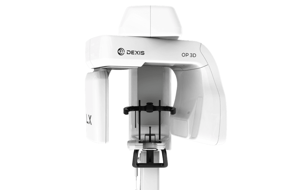
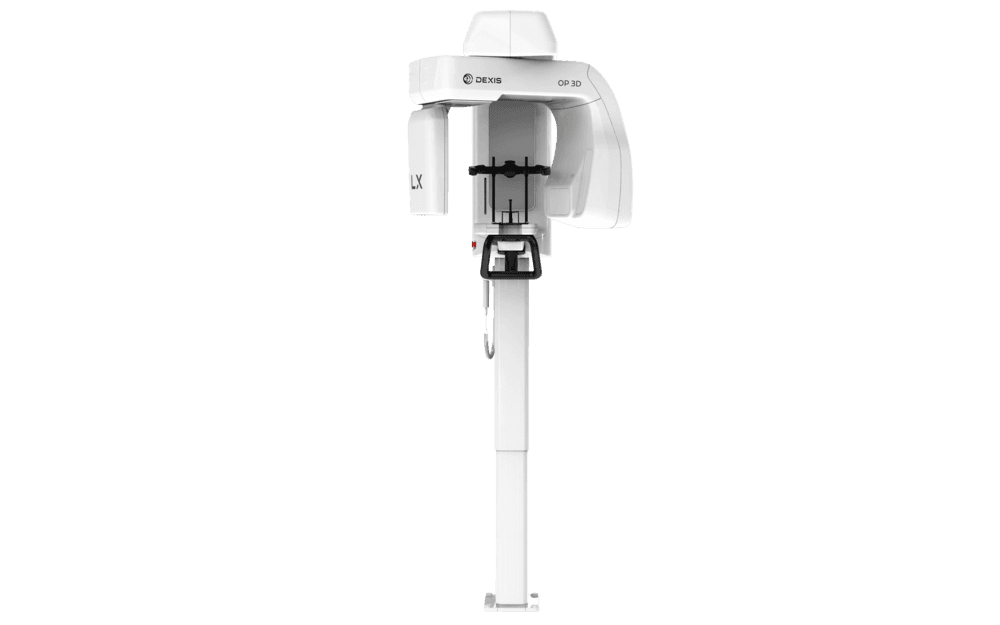
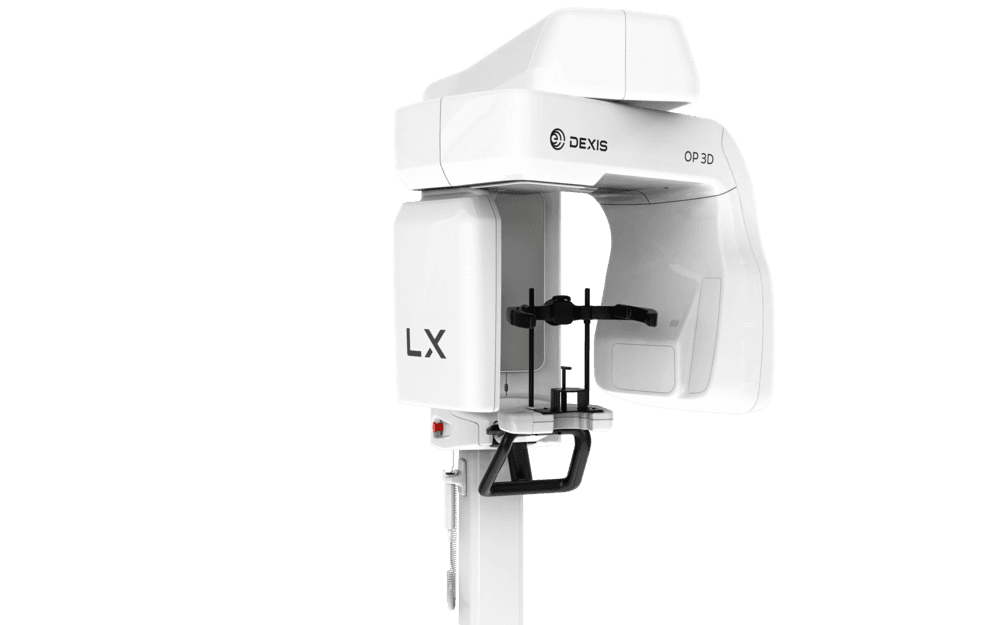
Dexis ORTHOPANTOMOGRAPH ™ OP 3D™ LX CBCT Unit
From 2D imaging to cephalometric and CBCT capabilities, the DEXIS Orthopantomograph OP 3D LX covers it all. It represents the next generation of DEXIS OP 3D technology, offering the largest sensor in the lineup, expansive field of view (FOV) options and a range of advanced, innovative features, including artifact and noise reduction, a new head support design and more.
Description
Imagine having all your 2D and 3D imaging needs met with a single device. That's exactly what the DEXIS Orthopantomograph OP 3D LX offers: a comprehensive 2D/3D imaging system that covers the full spectrum of dental extraoral applications — from endodontics and implant placement to airway analysis, orthodontics and more. Building upon DEXIS’ renowned OP 3D technology, the DEXIS OP 3D LX features the largest sensor in the lineup and offers flexible FOV options, ranging from 5x5 quadrant scans to 15x20 full maxillofacial complex high-resolution scans. Plus, with the ability to capture large diagnostic areas in a single, non-stitched scan — along with Cloud-based service connectivity — the OP 3D LX streamlines workflows, making them faster and more efficient.
- Artifact and noise reduction. The DEXIS OP 3D LX captures consistently clear, high-quality dental X-rays and CBCT scans, thanks to its innovative artifact and noise reduction filters
- Flexible FOV options. Comes standard with 2D panoramic imaging, extraoral bitewing and 3D CBCT capabilities with FOVs up to 12x15. Optional upgrades include expanding to a 15x20 volume size or adding cephalometric imaging capabilities, whichever you see fit
- Customizable user interface. Offers an intuitive user interface that easily lets you select your field of view, choose your imaging setting — 3D, panoramic or cephalometric — and make vertical and bi-directional scout adjustments
- Clear visibility. This DEXIS CBCT unit features automated ICE (Implant Contrast Enhancer) and MAR (Metal Artifact Reduction) technology to improve visibility of implants and reduce interference from metal and restorations
- Instant previews. Displays the dental X-ray image shortly after exposure, eliminating the need to open any image viewing software
- Advanced patient positioning. The DEXIS OP 3D LX is built with a newly reimagined head support design that captures scans without interfering with the patient's soft tissue profile, making it ideal for orthodontic and surgical applications
Specifications
Focal Spot: 0.5 (IEC 60336/2020)
Tube Voltage: 60 – 95 kV
Tube Current: 2 – 16 mA
HU Capacity: 35 kJ, 49 000 HU
Minimum Total Filtration: 3.4 mm Al @ 95 kV
Wheelchair Accessible: Yes
|
2D |
Panoramic |
Cephalometric |
|
Image Detector |
IGZO TFT |
IGZO TFT |
|
Sensor Pixel Size |
99 μm |
99 μm |
|
Image Pixel Size |
95 μm |
95 μm |
|
Scan/Exposure Time |
1.4 – 9.0 s |
6.9 – 11.3 s |
|
Image Field Height |
128.4 – 187.0 mm |
180.0 – 235.9 mm |
|
Imaging Programs |
Standard, segmented standard, pediatric, segmented pediatric, bitewing, Lateral TMJ |
Lateral, pediatric lateral, PA, carpus |
|
Weight |
120 kg / 265 lbs |
155 kg / 342 lbs |
|
3D |
CBCT |
|
Image Detector |
IGZO TFT |
|
Image Voxel Size |
80 – 400 μm |
|
Scan Time |
8 – 30 s |
|
Exposure Time |
0.9 – 19.4 s |
|
Image Volume Sizes (HxD) |
50x50, 60x90, 80x80, 100x100, 120x150, 150x200 mm |
|
DICOM Support |
Yes |
|
Min. Room Height |
2100 – 2455 mm |
Minimum System Requirements for 3D Acquisition Workstation
CPU (Processor): Intel Core i5, i7 or Xeon, 4-cores or more
GPU (Graphics Processing Unit): NVIDIA Quadro T1000 8GB, NVIDIA GeForce RTX 3050 8GB
RAM (Memory): 16 GB or more
Storage (Hard Disk): 1 TB or more
Network Gigabit Ethernet: 1000 Mbs/s
Operating System: Windows 11 Pro or Enterprise 64-bit, Windows 10 Pro or Enterprise 64-bit
Display: 1920 x 1080 (Full HD) resolution or higher
Other: OpenCL 1.1 support, DVD-ROM drive, Anti-virus software
Notes: Please refer to software and device installation manuals for detailed requirements.