
- Home
- Digital Imaging
- Extraoral Imaging
- J. MORITA Veraviewepocs 3D R100
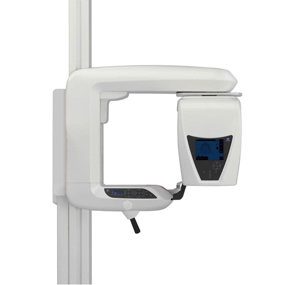
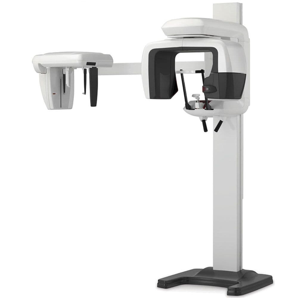
J. MORITA Veraviewepocs 3D R100
Description
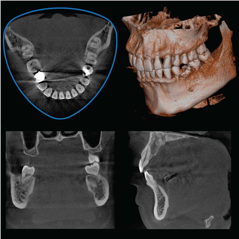
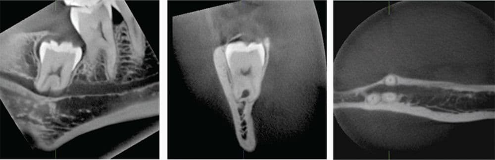
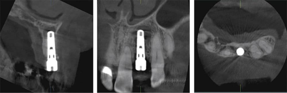
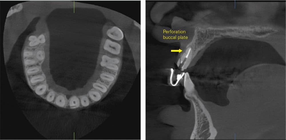
This unit’s groundbreaking 3D Reuleaux full arch field of view closely matches the shape of the natural dental arch. It reduces dose by excluding areas outside the region of interest and allows a complete scan of the maxilla and/or the mandible. 3D R100 is ideal for implantology as well as periodontology, orthodontics, endodontics, oral surgery, and general dentistry. It also offers TDO integration and compatibility with 3rd party imaging software with a DICOM export function. As with all MORITA units, it offers exceptional clarity and diagnostic capability.
- Multi-functional unit: 3D/Pan/Ceph
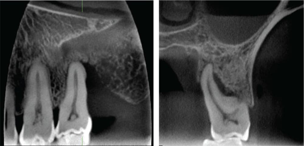
- Reveals bone structure, resorption, apical lesions, root fractures & more
- Six 3D fields of view
- Four options for easy & accurate 3D positioning
- Improved patient safety with Dose Reduction Mode
- New panoramic focal plane adjustment & partial pan/ceph image functions
- TDO Software integration with 1-click interface
- FREE 3-year warranty including remote software support
Specifications
Features:
- Model: X550
- 3D, panoramic, and cephalometric capabilities
- Super high resolution: 125 µm (micrometer) fine voxel
- Dose Reduction Feature lowers X-ray exposure 30% to 40%
- 3D exposure time: 9.4 seconds
- Panoramic exposure times: 7.4 seconds (standard); 15 seconds (high definition mode)
- FOV: Ø 40 x H 40 mm, Ø 40 x H 80 mm, Ø 80 mm x H 50 mm, Ø 80 mm x H 80 mm
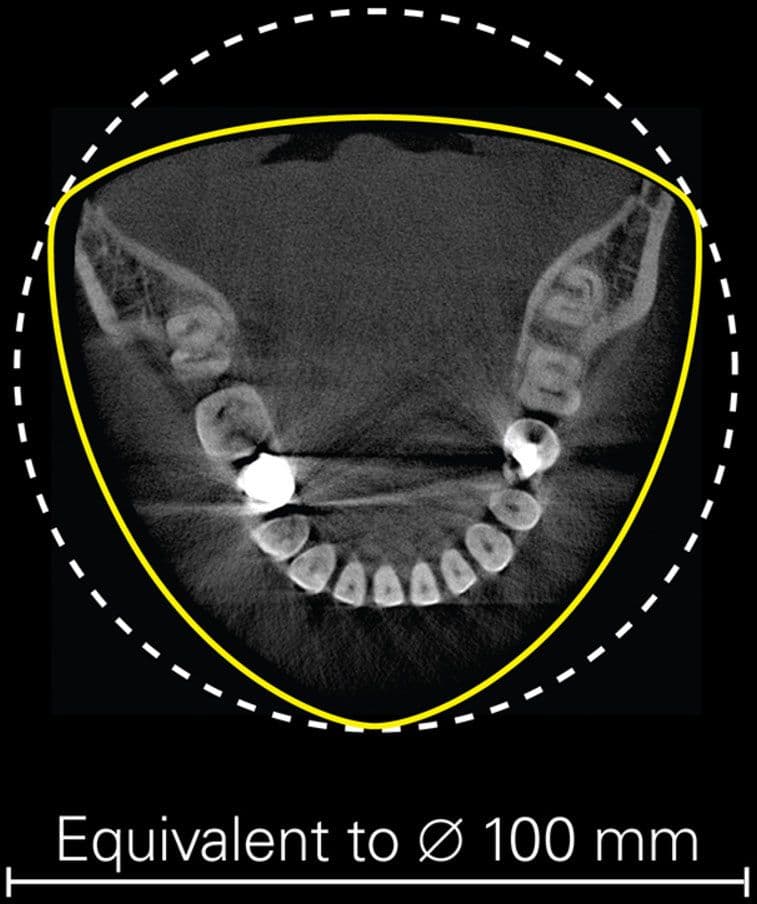
- 3D Reuleaux Full Arch FOV: Ø 100 mm (Equivalent) x H 50 mm, Ø 100 mm (Equivalent) x H 80 mm
- Dimensions: W 40.15" x D 51.18" x H 92.72" (without ceph)
- Dimensions: W 78.74" x D 51.18" x H 92.72" (with ceph)
- Weight: Approx. 419 lbs. / Approx. 573 lbs. with Cephalometric
- Tube voltage: 60-90kV (depending on exposure mode)
- Tube current: 1-10mA (depending on exposure mode)
- Input voltage: EX-1: AC 120V 60 Hz / EX-2: AC 220/230/240V 50/60 Hz
- Power Consumption: 2.3 kVA
- Effective focal spot: 0.5 mm
Imaging programs:
Standard Panoramic (standard, orthogonal and shadow reduction projections)
Magnification: 1.3 X throughout and 1.6 X throughout
Pedodontic Panoramic (standard, orthogonal and shadow reduction projections)
Magnification: 1.3 X throughout and 1.6 X throughout
Maxillary Sinus Panoramic (posterior and anterior)
Magnification: 1.5 X throughout
TMJ Quadruple Image
Magnification: 1.3 X throughout
Partial Panoramic
Magnification: 1.3 X throughout
Cephalometric image (option)
Projection: Posterior-anterior (PA) and Lateral (LA)