
- Home
- Digital Imaging
- Extraoral Imaging
- i-CAT™ FLX V-Series CBCT Unit
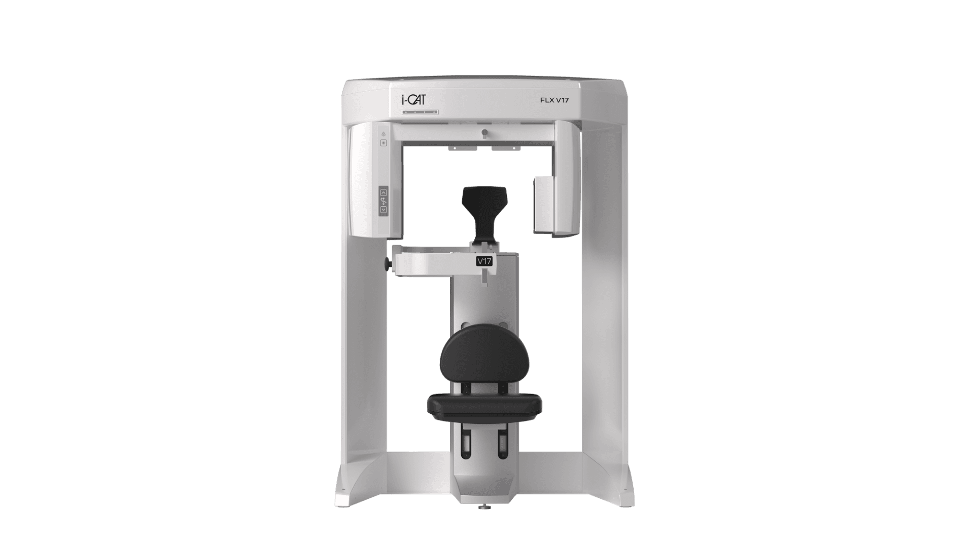
i-CAT™ FLX V-Series CBCT Unit
Description
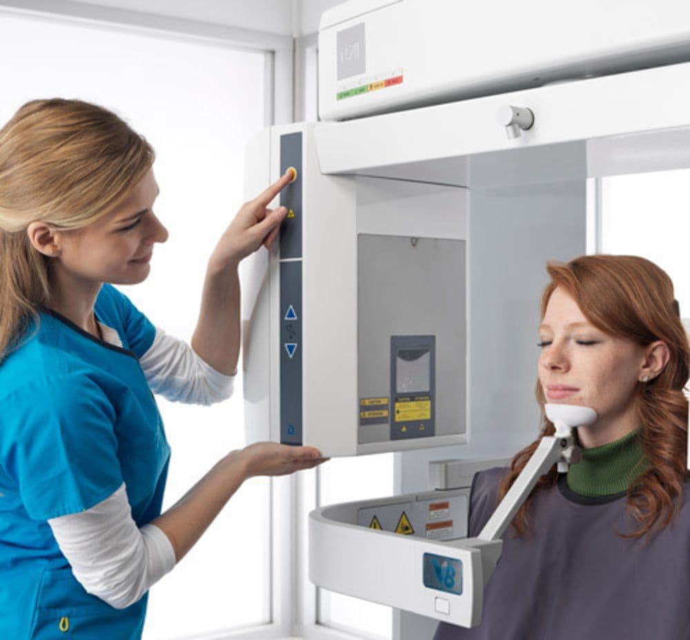
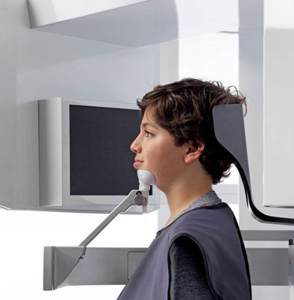
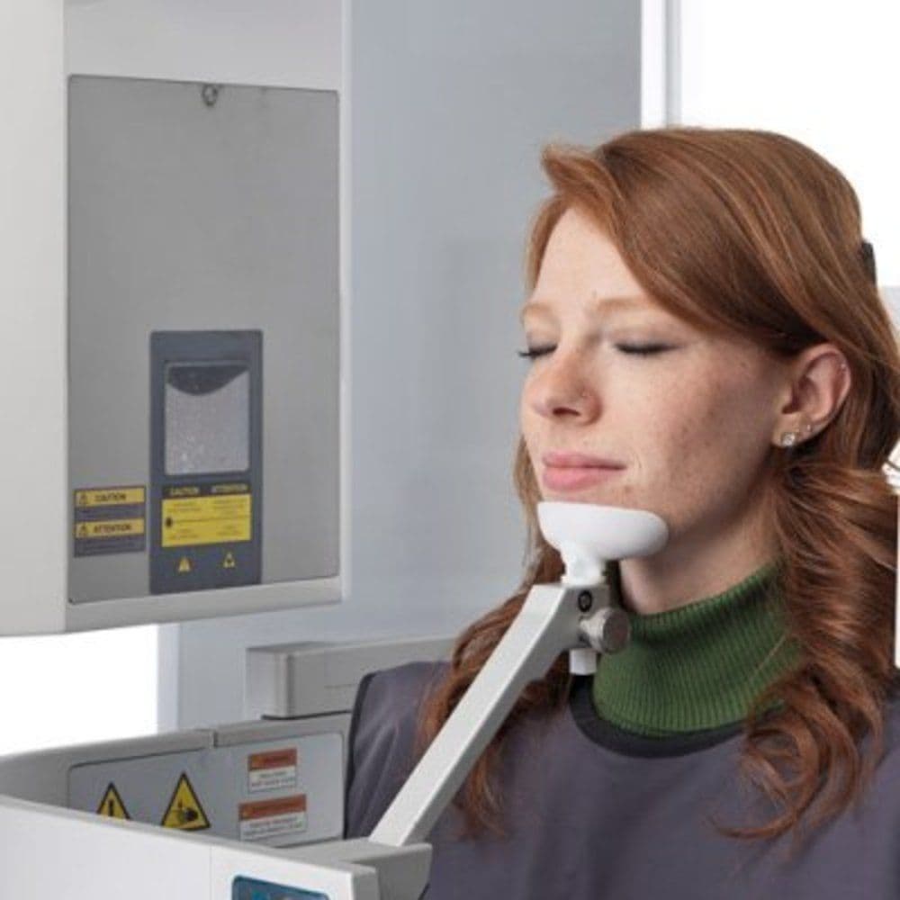
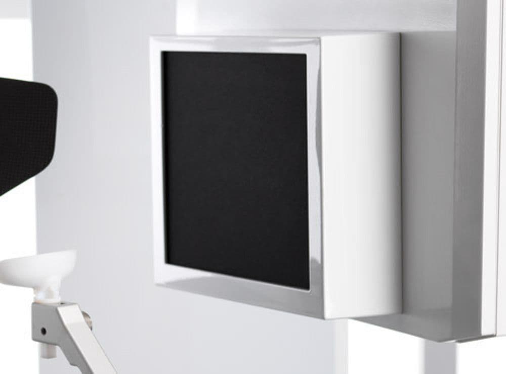
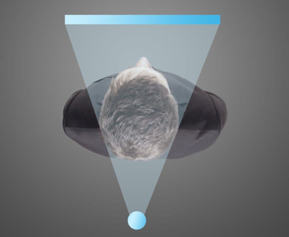
From enhanced low-dose 3D imaging options to traditional 2D panoramic capabilities, the i-CAT FLX V-Series CBCT unit is engineered to adapt to the needs of your practice, whether you’re new to digital dentistry or in the market for an upgrade. Available in three CBCT models — V8, V10 and V17 — the FLX V-Series comes equipped with full-beam scanning, maximizing the use of its large 24.2 cm x 19.3 cm sensor, without compromising image quality or anatomical accuracy. Plus, with a seated position built directly into its design, patients will naturally feel more comfortable — making them less prone to movement and unnecessary retakes.
- Flexible. Each CBCT machine is designed for a range of specialties and diagnostic capabilities, such as implantology, endodontics and oral maxillofacial surgery (all models), to more specific applications, such as TMJ (V10 and V17) and orthodontics (V17)
- Proactive. Leverages i-CAT QuickScan and QuickScan+ protocols to capture 3D images at comparable radiation doses to 2D panoramic images* — providing ultimate peace of mind
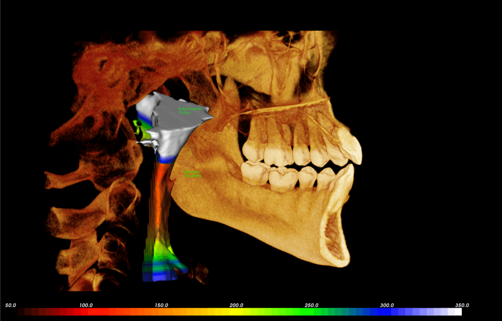
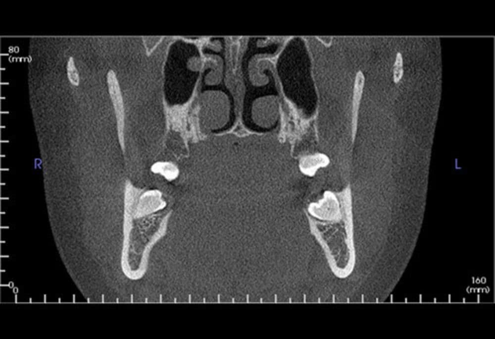
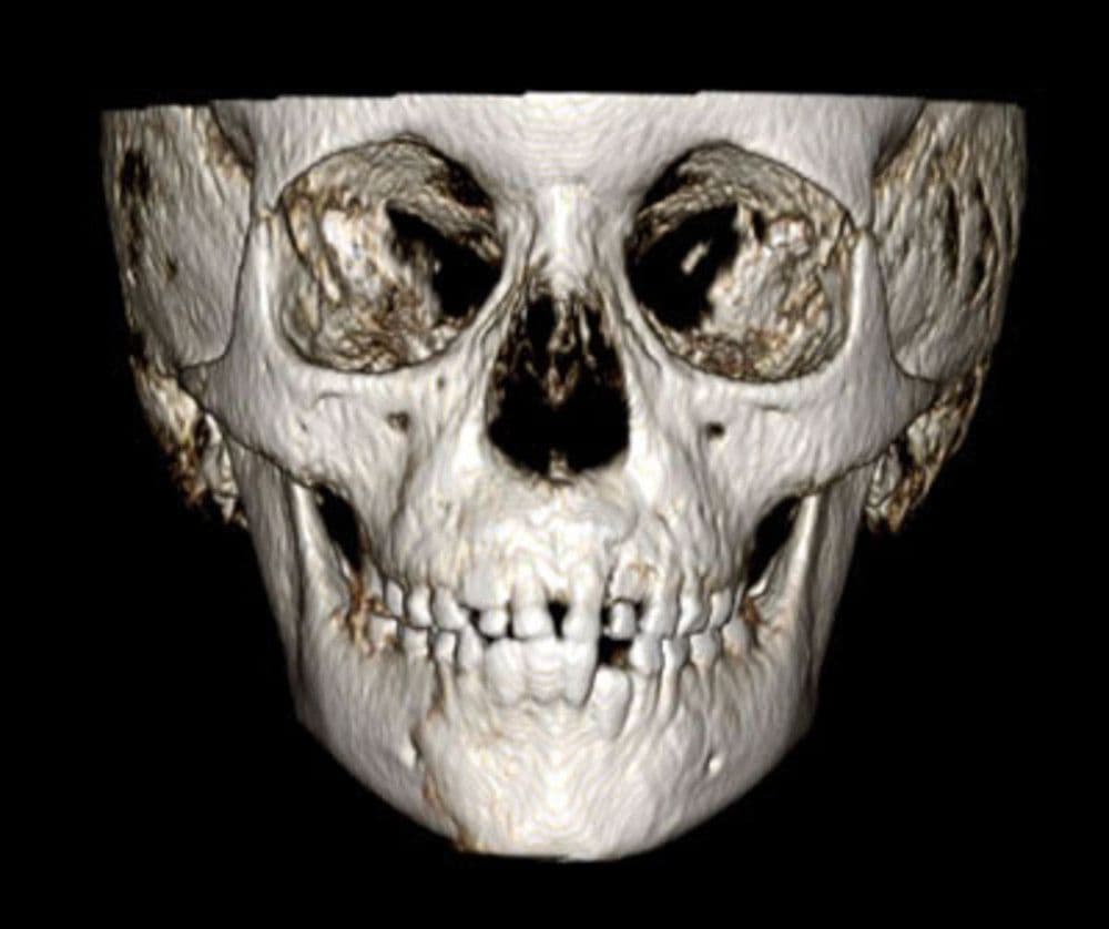
- Precise. Offers advanced digital imaging treatment planning tools, including high-resolution, volumetric images and complete 3D views, allowing for a more thorough analysis of bone structure and tooth orientation
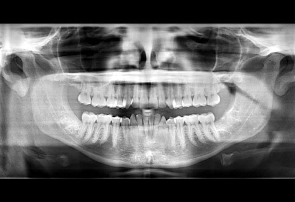
- Comprehensive. Takes traditional 2D pans using the same high-quality sensor as 3D images, for convenience that works at your own tempo
- Seamless. Encourages open platform compatibility with a variety of programs, such as CAD/CAM applications and other dental equipment
- Expansive. Available in a range of field of view options, including ø8x5cm to ø8x8cm (V8); ø8x5cm to ø16x10 cm (V10); and up to ø23x17 cm (V17)
* Utilizing the i-CAT FLX QuickScan+ exposure tool protocol. Use of lower dosage imaging may only be suitable for certain diagnostic tasks. Image quality is proportional to dose. i-CAT FLX offers a variety of exposure protocols allowing clinicians to adjust dosage to specific diagnostic needs.
FREQUENTLY ASKED QUESTIONS
1. How easy is it to upgrade the i-CAT FLX V-Series CBCT units?
Very easy. All that’s required is an in-office software upgrade that opens the field of view to the next (or desired) V-Series configuration — no hardware changes or downtime required.
2. What compatibilities are available?
Tx STUDIO, powered by Anatomage, is an integral part of the i-CAT CBCT workflow, combining the power of multiple software systems into one. Tx STUDIO can be used to aid in airway analysis and implant planning, as well as STL file export, surgical guides, face matching and more.
3. Which CBCT model is the most flexible?
Of the three CBCT models, the i-CAT FLX V17 CBCT unit offers the most flexibility for all your 3D imaging needs, with a maximum and scalable field of view of up to ø23x17 cm. It is a popular choice among orthodontists, oral maxillofacial surgeons and oral radiologists, but can be appropriate for any clinician seeking a full view of the entire oral-facial complex.
4. What are the other CBCT models best suitable for?
The V8 is suitable for implants, endodontics and most general dentistry, while the V10 is suitable for implants, periodontics, prosthodontics, airway assessment, TMJ and most oral and maxillofacial surgeries.
Specifications
- Voxel Size: .4 mm, .3 mm, .25 mm, .2 mm, .125 mm
- Collimation: Electronically controlled, fully adjustable collimation
- Scan Time: 4.8, 8.9, 14.7, 17.8, or 26.9 seconds
- Field-of-View:
V8: 8cm x 5cm, 8cm x 8cm
V10: 8cm x 5 cm, 8cm x 8cm, 16cm x 4cm, 16cm x 6cm, 16cm x 8cm, 16cm x 10cm
V17: 8cm x 5cm, 8cm x 8cm, 16 cm x 4cm, 16cm x 6cm, 16cm x 8cm, 16cm x 10cm, 16cm x 11cm, 16cm x 13cm, 23cm x 17cm - Typical Reconstruction Time: Less than 30 seconds
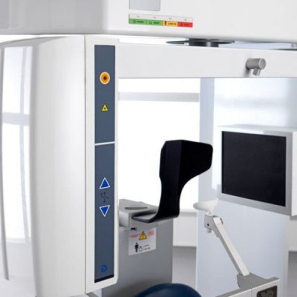
- Patient Position: Seated
- Unit Size: 48" (w) x 69.5" (h) x 36.37 (d)