
- Home
- Digital Imaging
- Extraoral Imaging
- DEXIS ORTHOPANTOMOGRAPH™ OP 3D™ CBCT Unit
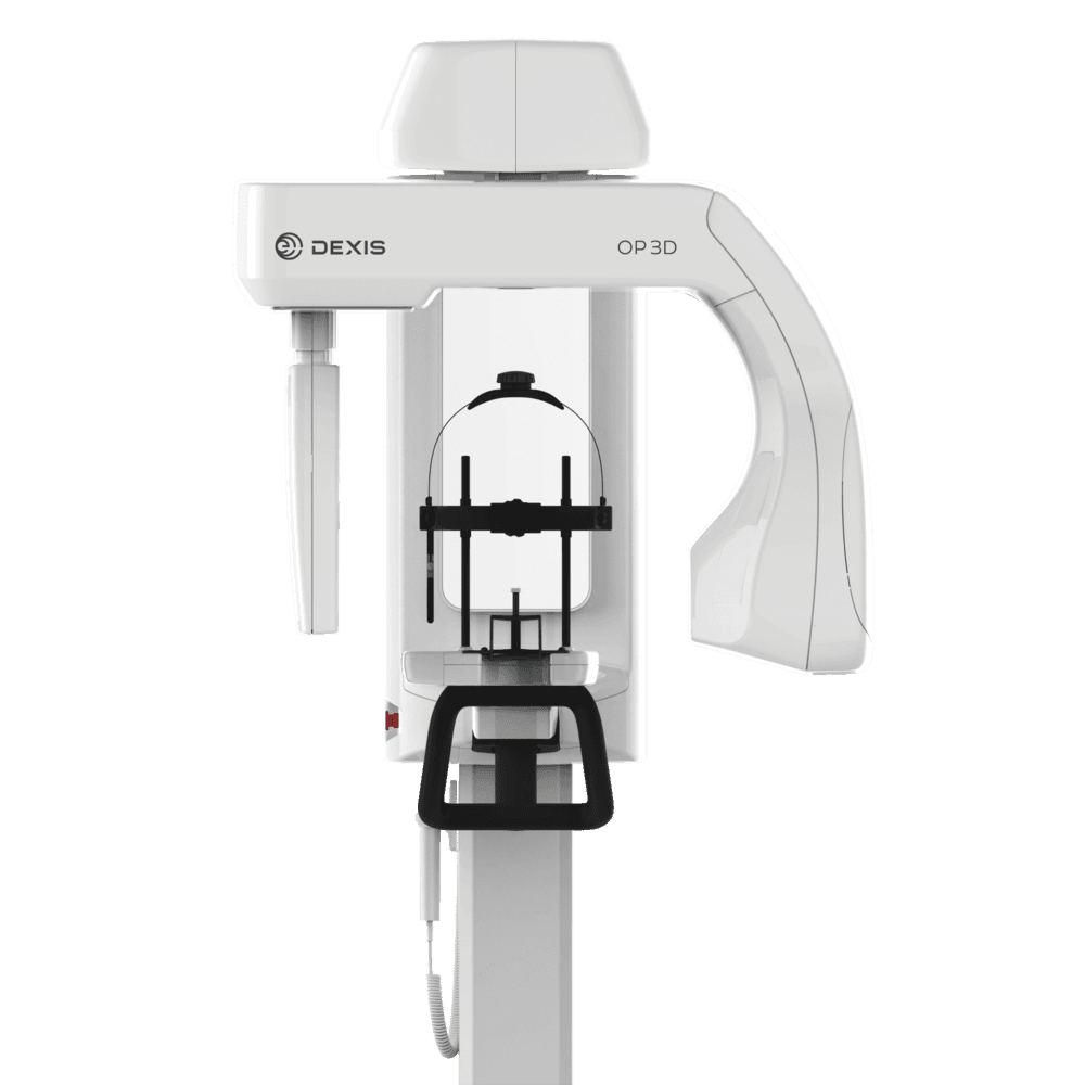
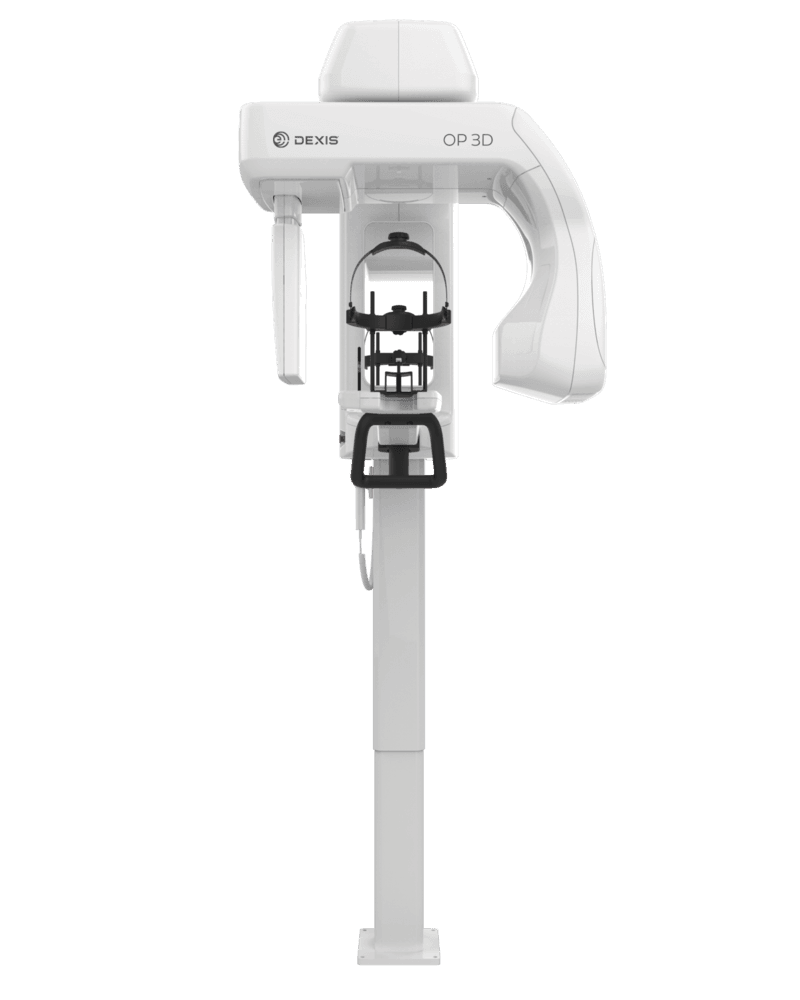
DEXIS ORTHOPANTOMOGRAPH™ OP 3D™ CBCT Unit
Description
Experience the transformative benefits of digital dentistry. The DEXIS OP 3D CBCT unit offers diagnostic precision, with various configurations — from panoramic-only through cephalometric and 3D imaging — that cater to a range of specialties and applications, including endodontics, implantology, TMJ and more. Equipped with an intuitive interface for easy operation, and an overall design that’s meant to be upgradeable and expandable, this innovative, lead-free CBCT machine offers advanced digital imaging capabilities that can grow and keep pace with your practice.
- User-friendly. Allows the exact imaging area to be selected, such as an individual tooth, an entire upper/lower jaw or the TMJ region, and automatically selects the ideal field of view for that selection
- Consistent. Delivers consistently sharp panoramic images — thanks to its built-in DEXIS ORTHOfocus™ feature — enabling forgiving patient positioning
- Fast. Offers a quick preview of the captured image for fast and efficient evaluation
- Simple. Keeps workflows simple by allowing all non-patient positioning functions to be easily and intuitively controlled via laptop or PC
- Configurable. Available in panoramic, cephalometric and 3D imaging configurations, with field of view options based on true clinical needs: ø5x5 cm, ø6x9 cm, ø9x11 cm and ø9x14 cm (optional)
FREQUENTLY ASKED QUESTIONS
1. What are the differences in the field of view sizes?
The DEXIS OP 3D CBCT unit offers a variety of configurations to meet your precise field of view needs:
- ø5x5 cm: optimized for single-tooth and localized diagnostics
- ø6x9 cm: offers the ability to scan either the lower or upper jaw
- ø9x11 cm: covers the entire dentition, including the lower and upper jaw and part of the maxillary sinus
- ø9x14 cm: the largest configuration available, ideal for capturing the entire craniofacial area
2. What are the DEXIS OP 3D CBCT unit’s panoramic imaging capabilities?
This CBCT machine offers a variety of panoramic-specific programs, including:
- Pediatric panoramic: features a clinically adapted image layer and reduced image height
- Bitewing: provides a quick and easy alternative to intraoral bitewing imaging
- TMJ: provides a lateral view of the temporomandibular joints, with an open or closed mouth
3. What are the DEXIS OP 3D CBCT unit’s cephalometric imaging capabilities?
The DEXIS OP 3D CBCT unit offers comprehensive cephalometric imaging capabilities, including:
- Lateral cephalometric images: enables rich anatomical details with exceptional visibility of the soft tissue borderline; ideal for adult and pediatric patients
- Pediatric lateral images: features reduced height and minimal radiation exposure for dose-sensitive pediatric patients
- PA cephalometric images: produces excellent details, thanks to its powerful dedicated X-ray source
- Carpus images (optional): gathers vital information for determining patient age and growth
4. What is the QUICKcompose™ feature?
The DEXIS OP 3D CBCT unit is equipped with an intuitive feature called QUICKcompose. This allows a preview of a captured image to be displayed on the user interface as soon as the scan is completed, and is available in panoramic, cephalometric and 3D imaging configurations.
Specifications
• Image Volume Sizes (HxW): 5 x 5, 6 x 9, 9 x 11, optional 9 x 14 cm
• Imaging Programs: Standard, Segmented, Pediatric,Lat TMJ, Bitewing
• Image Voxel Size: 80 μm-400 μm
• Image Detector: CMOS
• Scan (2D): 9 s
• Scan Time (3D): 10-20 s
Technical Details
|
Focal Spot |
0.5 mm, IEC 336 (IEC 60336/2005) |
|
Tube Voltage |
60-95 kV |
|
Tube Current |
3.2–16 mA |
|
2D Panoramic |
|
|
Image Detector |
CMOS |
|
Sensor Pixel Size |
99 µm |
|
Image Pixel Size |
99 µm |
|
Scan/Exposure Time |
9 s |
|
Image Field Height |
147 mm |
|
Imaging Programs |
Standard, Segmented, Pediatric, Lat TMJ, Bitewing |
|
Wheelchair Accessibility |
Yes |
|
3D |
|
|
Image Detector |
CMOS |
|
Image Voxel Size |
80 µm-400 µm |
|
Scan Time |
10–20 s |
|
Image Volume Sizes (H xW) |
5 x 5, 6 x 9, 9 x 11, 9 x 14 cm |
|
DICOM Support |
Yes |