Dr. Anthony Lavacca, DMD, FACP, FICOI
A certain synergy takes place between clinicians in a multispecialty office. My practice, Naperville Dental Specialists, offers cosmetic and functional services for all ages, so my patients can stay with me and my team for all of their dental needs. My wife, Dr. Manal Ibrahim of Innovative Orthodontic Centers, and I can treat patients as single specialists or as a team. It is important that we have dental equipment that helps us gain a more precise diagnosis for the best results. This is definitely the case with the i-CAT™ FLX™ Cone Beam 3D imaging system.
3D imaging—more precise diagnosis to plan for the best results
i-CAT scans can be used for a wide variety of procedures and shared with referring doctors. Patients can benefit from 3D scans as well as traditional 2D radiography. When an X-ray is needed, I can acquire a traditional pan or a low-radiation dose scan from the i-CAT FLX.
The pan view can be pulled from the 3D scan. The scan data is extremely valuable when planning for surgical extractions. If we see a sign of caries, we can take an individual bitewing or periapical with our DEXIS™ Platinum sensor. This intraoral digital imaging is also useful intra-surgically when placing implants.
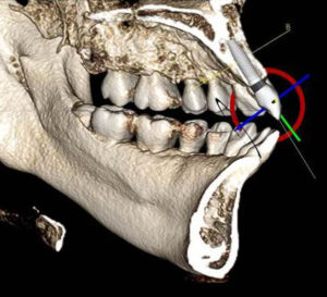
As a prosthodontist and an implantologist, I’ve found the anatomical information from a 3D scan helps me place implants more accurately and efficiently. With the 3D view, I can achieve a greater understanding of the shape, width, and height of the bone and surrounding structures prior to surgery. Tx STUDIO™ software that comes with an i-CAT system gives me access to integrated treatment tools for implant planning and the ability to integrate use of surgical guides. This reduces significantly the time it takes to place the implant and translates into less discomfort and healing time for the patient.
With orthodontist-prosthodontist Dr. Ibrahim, I can preplan potential implant sites during orthodontic treatment and capture all of our initial imaging needs in a single scan, with accurate 3D views of the teeth, roots, TMJ, airway, and sinuses. Dr. Ibrahim can evaluate tooth position and see impacted teeth and supernumeraries while I explore the need for bone grafting. During the combination of implant and orthodontic treatment, she can move teeth to the appropriate position, and by the time orthodontic phase is complete, the bone graft is ready for the implant area. Once implants are placed, we can capture a low-dose QuickScan+, a setting that gives us a full-dentition 3D scan at a lower dose than a panoramic X-ray*, to check if the teeth have moved into proper position in the bone.
From a highly visual 3D scan, patients can see where the teeth must move, where we will position the implants, and how all the treatment will turn our plans into reality.
*Data on file with i-CAT
Dr. LaVacca has no financial interests in i-CAT.
About the Author: Dr. Anthony LaVacca received a Doctor of Dental Medicine degree from Temple University. He received post‐graduate education in prosthodontics from Montefiore Medical Center/Albert Einstein College of Medicine. Dr. LaVacca is an American Board‐certified prosthodontist and fellow of the American College of Prosthodontists. At the Montefiore Medical Center/Albert Einstein College of Medicine in Bronx, NY, Dr. LaVacca served in several positions, including director of the general practice residency program, interim director of the postgraduate program in prosthodontics, and assistant professor. He is currently practicing at Naperville Dental Specialists & General Oral Health Care in Naperville, Illinois.