It must meet the challenges and satisfy the clinical demands of your practice, your patients and lab partners. It is my opinion that digital impressioning has achieved the status of the gold standard in restorative dentistry. Computers operate with an efficiency and an accuracy that conventional materials cannot consistently attain. One such technology that truly meets these criteria is the 3M™ ESPE™ Lava Chairside Oral Scanner C.O.S. The following case illustrates how the Lava C.O.S. can provide excellent results in restorative dentistry from impressioning to seating.
Case Report
The patient, a 79-year-old female, presented to the office with a failed fixed partial denture #2XX5. The prosthesis had failed due to recurrent caries beneath the mesial abutment. The patient was given all options for replacement of the failed restoration. She chose to have us fabricate a new ceramometal fixed-partial denture.
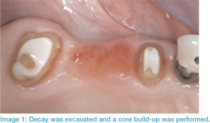
Following accepted restorative protocols, the old prosthesis was removed and all decay beneath the abutments was excavated. FluoroCore® (Dentsply, Inc.) was used as a foundation for the preparations, which were then refined to support a four-unit fixed-partial denture. Its design included a semiprecision slot connection between the pontic #4 and abutment tooth #5.
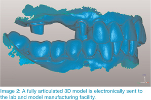
We then proceeded to isolate the teeth in order to scan the full arch using the Lava C.O.S. After verifying that 100% of the preparations were captured in the scan, the jaw relation was recorded by the scanner with the patient closed into centric occlusion. A provisional restoration was fabricated and the patient was dismissed.
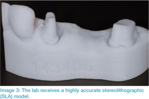
The digital information, along with a digital prescription, was transferred wirelessly to the Authorized Lava Design Center. The lab used the digital files to mark the margins. The virtual model was then sent to a model-manufacturing facility to produce a stereolithographic (SLA) model.
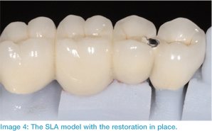
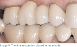
This model was used for fabrication of the prosthesis, using the same techniques that would be followed if using gypsum die stone. (The difference is that the digital information does not change dimension in any way after it is captured by the scanner. The same cannot be said for conventional impression materials.) We placed the final restoration without any adjustments needed. The patient was ecstatic about the complete treatment process and she was truly amazed at the difference this new technology brings.
Placing restorations without making any adjustments has become the norm since incorporating the Lava C.O.S. The fit of the restorations is so accurate that rarely are any adjustments needed, provided that the provisional restoration satisfies all requirements.
The Benefits of Digital
When implementing the Lava C.O.S. in my practice, my goal was to provide an increased benefit to the patient through delivering a higher quality restoration. In the end, the benefits of the Lava C.O.S. have extended to my practice as well.
Dentists who have used the system reported a 41% reduction in seating times for single-unit crowns1 and remake rates due to marginal fit were up to 80% lower than the industry average.
In fact, since using the system in my practice, only 6 units of 250 have required modification prior to cementation. None of the errors were related to the scanner. My experience with the first 250 units scanned has been exceptional from both the perspective of my patients and my own. With experience and with continued software improvements from 3M, I see no tarnish on this technology.