Three-dimensional radiographs provide precise data that dentists can use to make informed choices for each patient's individualized procedure and each treatment plan. The i-CAT offers many options in Cone Beam Computed Tomography (CBCT) scans to help dentists to approach innovative procedures with confidence.
Flexibility in scan sizes:
The i-CAT has a great amount of flexibility in imaging selections that help to reduce radiation. Practitioners have the ability to choose the size of scan appropriate for various treatments, while reducing radiation exposure and helping them to achieve their ALARA goals. The system's unique feature with the full field-of-view of 16-cm diameter and offers
and extended field-of-view option of 17cm x 23 cm. The system's feature of collimation, or sizing, facilitates a targeted view of the region of interest by allowing the clinician to change the height from 2 cm to 13 cm or anything between. Furthermore, this collimation happens at the radiation source, thus the reduction of exposure as the scan size lessens.
Flexibility in scan times:
Scan time relates to resolution and exposure, and the i-CAT offers several choices. The popular 5-second scan significantly lowers emitted dose, especially useful when follow up scans are deemed necessary.
Flexibility for treatment planning:
The extended field of view allows for a complete orthodontic workup, frontal and lateral cephalometric, panoramic, supernumerary, SMV, TMJ, and airway studies. For those who have expanded their practice to include implants, the scans optimize implant placement and selection of the most suitable implant type, size, location, and angulations prior to
surgery. i-CAT scans are also easily transferred into most major orthodontic and implant planning software.
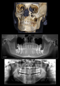
Flexibility in dimensions:
For those who require a combination of 2-D and 3-D imaging, the i-CAT has you covered. The system offers both i-PAN, a traditional 2-D pan function and Tru-Pan™ that delivers precise panoramic views from 3-D scans (reconstructed pan).
Flexibility in viewing options:
i-CAT images can be enlarged, rotated, and sliced in any direction. They can be viewed with or without Quantum IQ™ image enhancement within the included i-CATVision™ 3-D imaging software. Plus, this software and the patient's scan can be shared with colleagues so they too, have these viewing options.
Flexibility in scheduling:
Having an in-office cone beam scan exposes the patient to less radiation than a medical CT scan. Besides, with an in-office scanner, the patient does not need to make additional appointments for imaging, and then for treatment planning after scanning. The i-CAT offers the fastest time from scan-to-plan to consult—about one minute from scan capture to the images showing in the consultation room or operatory.