CariVu — No radiation, 99% accuracy, and software integration
The DEXIS™ CariVu™ caries detection device helps me diagnose caries with great accuracy and without exposing patients to ionizing radiation.
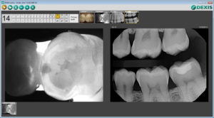
It uses transillumination technology – the image on the screen is similar to that of an X-ray, and the enamel appears transparent while porous lesions appear as dark areas. This is especially helpful for the identification of occlusal, interproximal, and recurrent carious lesions and cracks not visible on other imaging methods.
CariVu fulfills four very important niches in my dental practice.
Imaging without ionizing radiation.
Some patients are reluctant or unable to be exposed to radiation: pregnant patients, young children, people with medical conditions for which they have already been exposed to radiation, and patients who have recently received dental X-rays. Taking a “full-mouth series” with a CariVu can offer important information on caries development.
A “double check” for carious lesions and cracks unclear on X-rays.
When a radiolucency on a radiograph is not clear enough for a definitive diagnosis, or if an incipient lesion goes around the interproximal and curves toward the lingual or buccal, a CariVu image offers a big advantage.
Detecting caries at an earlier stage.
Without CariVu, I put a “watch” on that patient’s chart or take an “educated guess” if I see a “darkness” in the interproximal area of the tooth. I would rather have a transilluminated image that shows the carious area with a 99% accuracy* rate than take the chance that the caries may reach the pulp before the patient’s next appointment, or infected structure may be lurking beneath a carious lesion. The CariVu image also shows me if a certain spot can, in fact, just be watched and given a course of preventative care before treatment. During cleaning, the reparative dentin can cause staining sometimes mistaken for caries, and CariVu is an efficient double check for that situation as well.
Patient education.
Patients have an easier time identifying a problem on a transilluminated image. My team and I also appreciate that we can get images on children whose small mouths or gag reflexes make radiographs difficult.
CariVu images are captured from the portable handpiece and go directly into the DEXIS software to be stored with the patient’s intraoral X-rays and camera images.
It has increased the objectivity, accuracy, and safety of my diagnostic process. It’s also contributed to better communication with my patients.
Originally published in Sidekick Magazine
*Kühnisch J. Benefits of the DIAGNOcam Procedure for the Detection and Diagnosis of Caries [study project]. Munich: Ludwig Maximilian University of Munich; 2013. Indications for use are found at www.dexis.com/ifu.
About the Author: Dr. Tigran Khachatryan earned his dental degree from the University of Washington School of Dentistry. He is a Kois Center graduate and practices in Redmond, Washington at A Smiling Heart Dentistry. Dr. Khachatryan has no financial interests in DEXIS, LLC.