When my wife and I were naming my dental practice, we wanted it to represent our desire to reach beyond the physical smiles of clinical dentistry because a person is more than their physical appearance. A Smiling Heart Dentistry was born to bring “Smiles from Your Heart.” To comprehensively treat the person, we utilize many technologies, including 3D imaging.
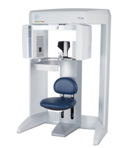
To place implants responsibly and confidently, I need precise information about my patient. When I started offering implants, I would send patients next door for a 3D CBCT scan. When we moved, the technology plan for my new office included an in-house 3D imaging system, an i-CAT FLX.
 3D imaging is essential for a practitioner who regularly places implants. Information can be obtained regarding the bone measurements, nerve location, and sinuses, to name a few aspects of the critical anatomy. With “old fashioned” dentistry techniques, the clinician can try to “feel” the amount of bone available for an implant with a caliper. Even with a 2D X-ray modality, it can appear there is sufficient bone to place an implant when, in reality, there may only be a couple of millimeters of bone. These detailed measurements of bone height and density can be quickly obtained from a 3D scan.
I researched many types of CBCT units. I am a big proponent of exposing my patients to the least amount of radiation possible. For this reason, I chose i-CAT FLX because I can capture a 3D image with a dose lower than a 2D panoramic X-ray on the QuickScan+ setting. If I need to ensure all aspects of the implant are in the proper position, I can capture a post-treatment, low-dose image and still be within a reasonable exposure range.
Using 3D scans keeps the patient involved since I can better educate them in the diagnosis and treatment planning phases. My ability to rotate the 3D volume and virtually place the implants helps them understand their dental condition — a huge plus.
Besides implants, having a 3D imaging method has allowed me to expand procedure options. The software tools help me gather information for those suffering with airway obstructions or other airway disorders to recommend further diagnostic procedures and proper treatment. We can also gain information about the TMJ, and I can convert the 3D into a lateral ceph for orthodontic purposes.
If I see an area of concern, I can electronically send the scan securely to any specialist or colleague in the world with the click of a button. And, it’s the same for sending the scan to a lab or to fabricate a surgical guide.
I like that out of just one scan, I can create a panoramic, a lateral ceph, and see soft tissue and sinuses. I can virtually place implants in front of the patient, even virtually putting on skin with 3D imaging software. This level of pre-treatment planning blows the patients away and gives them confidence. The mission of A Smiling Heart Dentistry is to produce smiles on the patients’ faces and in their hearts, and if that can be accomplished, my team and I will be smiling as well.
Originally published in Sidekick Magazine
About the Author: Tigran Khachatryan DDS, obtained his dental degree from the University of Washington School of Dentistry. He is a Kois Center graduate and practices in Redmond, Washington at A Smiling Heart Dentistry.
